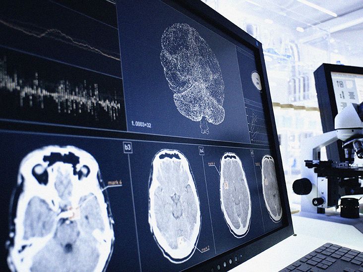
For decades, biology textbooks treated the brain like a celebrity hiding behind velvet ropes: important, delicate, and largely separated from the immune crowd. The blood-brain barrier seemed to keep most immune activity outside, while the central nervous system enjoyed a special status called “immune privilege.” That picture was useful, but incomplete.
Researchers now understand the brain as protected rather than isolated. It has resident immune cells, guarded borders, fluid-drainage routes, and constant communication with immune organs throughout the body. The system is less like a locked bunker and more like a selective international airport: approved cargo gets through, suspicious signals trigger screening, and occasionally security causes a bigger commotion than the original problem.
This surveillance protects neurons from infection, clears damaged material, helps shape neural circuits, and coordinates healing. Yet the same machinery can contribute to multiple sclerosis, autoimmune encephalitis, stroke damage, Alzheimer’s disease, and other neurological disorders when it becomes overactive or fails to switch off.
The Brain Is Immune-Privileged, Not Immune-Free
“Immune privilege” does not mean the immune system ignores the brain. It means immune responses in the central nervous system are tightly controlled because neurons are difficult to replace and inflammation can disrupt delicate electrical networks. A swollen ankle is unpleasant. Swelling inside a rigid skull is an entirely different level of bad planning.
The brain relies on overlapping defenses. The blood-brain barrier controls access from the bloodstream. Microglia patrol brain tissue. Immune cells in the meninges monitor its borders. Cerebrospinal fluid carries molecular information, while meningeal lymphatic vessels drain fluid and brain-derived material toward lymph nodes. Together, these systems watch without constantly storming the control room.
The Blood-Brain Barrier: Gatekeeper and Signal Station
A Selective Filter, Not a Brick Wall
The blood-brain barrier, or BBB, is formed mainly by tightly connected endothelial cells lining the brain’s small blood vessels. Pericytes and the end-feet of astrocytes help maintain this specialized environment. Tight junctions limit uncontrolled movement between blood and brain, while transport proteins admit glucose, amino acids, and other essentials.
This arrangement blocks many pathogens, toxins, inflammatory molecules, and circulating immune cells. It also makes drug development difficult: a medicine can look brilliant in a laboratory dish and then arrive at the BBB without the correct invitation.
The Barrier Listens to the Body
The BBB does more than stand guard. Its cells detect immune messengers in the bloodstream and relay information into the central nervous system. In a 2026 mouse study involving West Nile virus, viral signals in a distant part of the body activated antiviral genes in brain blood-vessel cells within hours, before the virus was detectable in the brain. The barrier appeared to serve as an early-warning station, preparing microglia and other defenses for a possible attack.
During severe infection, trauma, stroke, or prolonged inflammation, the BBB may become more permeable. Antibodies, inflammatory proteins, and immune cells can then reach areas normally closed to them. Sometimes that access helps fight disease; sometimes it adds fuel to the fire.
Microglia: The Brain’s Resident Patrol
Microglia are the primary resident immune cells within brain tissue. Their branching processes continuously sample the surrounding environment, checking for damaged membranes, abnormal proteins, dying cells, infection, and changes in neuronal activity.
When microglia detect trouble, they move toward the affected area, release signals, engulf debris, and attack certain pathogens. This cellular swallowing process, called phagocytosis, makes them part security team, part sanitation crew, and part neighborhood watchexcept the neighborhood contains roughly everything you have ever learned.
They Also Maintain Brain Circuits
During development, microglia help remove unnecessary synapses so neural circuits become more efficient. In adulthood, they support synaptic health and release factors that influence repair and neuronal survival. Their behavior varies with age, brain region, genetics, metabolism, and the type of threat. A response that clears debris after an injury can become harmful if it persists for months or years.
The Meninges: An Immune Command Post at the Border
The brain and spinal cord are wrapped in three membranes called the meninges. Once discussed mostly as protective packaging, these layers are now recognized as active immune environments containing macrophages, dendritic cells, T cells, B cells, and other specialized populations.
Many of these cells can monitor cerebrospinal fluid and blood-derived signals without sending a large inflammatory force directly among neurons. They collect antigensmolecular clues from damaged tissue or possible invadersand help determine whether a broader adaptive immune response is needed.
This border strategy lets the immune system inspect what is leaving the central nervous system and communicate with lymph nodes while keeping routine immune traffic away from sensitive neural circuits.
Meningeal Lymphatic Vessels: Drainage and Reporting
A major shift in neuroscience occurred when researchers demonstrated functional lymphatic vessels in the meninges. These vessels run near major blood vessels and connect with deep cervical lymph nodes in the neck. Their location helps explain why they remained hidden for so long: biology tucked important plumbing behind other plumbing.
Meningeal lymphatic vessels drain cerebrospinal fluid, immune cells, and brain-derived molecules. Material from brain tissue can move through fluid-clearance pathways toward the meninges, where lymphatic vessels provide an exit toward peripheral immune organs.
This route supports surveillance because lymph nodes are places where immune cells inspect antigens and coordinate adaptive responses. However, the same communication may participate in autoimmune disease when the immune system incorrectly targets nervous-system components.
How a Brain Immune Response Unfolds
Although every disease follows its own script, brain immune surveillance often follows a recognizable sequence:
- Detection: Microglia, barrier cells, astrocytes, neurons, or border immune cells sense pathogens, damaged cells, or inflammatory signals.
- Communication: Cytokines, chemokines, and interferons alert nearby cells and recruit help.
- Containment: Barrier cells strengthen antiviral defenses, while local immune cells gather around damage.
- Cleanup: Microglia and macrophages engulf dead cells, damaged myelin, abnormal proteins, and debris.
- Resolution: Repair programs should reduce inflammation after the danger passes.
The final step is crucial. Inflammation is not automatically harmful; uncontrolled or unresolved inflammation is the larger concern. A response that refuses to leave is like a fire department extinguishing the kitchen blaze and then flooding the house for six months.
When Protection Turns Into Damage
Multiple Sclerosis and Autoimmune Encephalitis
In multiple sclerosis, immune cells attack myelin, the insulation around nerve fibers, disrupting communication between the brain and body. In autoimmune encephalitis, antibodies or immune cells mistakenly target brain proteins, sometimes including receptors involved in neuronal signaling. Symptoms can include memory problems, seizures, unusual movements, psychiatric changes, confusion, and altered consciousness. Suspected encephalitis requires urgent medical evaluation.
Neurodegeneration, Infection, and Injury
In Alzheimer’s disease and related disorders, microglia may initially clear abnormal proteins and injured cells. Aging, genetics, and persistent debris can later push them toward dysfunctional inflammatory states. After infection, stroke, or head trauma, immune cells also clear damage and support repair, but excessive inflammation can enlarge injury. Older immune cells may have more difficulty removing debris and resolving the response.
The Brain and Body Keep Talking
Signals travel in both directions. The brain influences immunity through the autonomic nervous system, stress hormones, sleep, behavior, and endocrine pathways. Immune molecules produced during illness can alter body temperature, appetite, energy, mood, pain sensitivity, and attention.
This conversation helps explain why an infection in the body can make thinking feel slower even when the pathogen never enters the brain. It also guides research into how gut inflammation, chronic disease, stress, and aging affect neurological health. Animal studies have identified striking pathways, but those findings must be translated cautiously before being applied broadly to people.
What Researchers Hope to Do Next
The goal of neuroimmunology is not simply to “boost” or “suppress” immunity. Researchers want to tune particular cells and signals at the right location and disease stage.
Strategies under study include restoring healthier microglial metabolism, encouraging protective microglial states, improving lymphatic drainage, strengthening or temporarily opening the BBB for targeted drug delivery, blocking harmful antibodies, and limiting specific inflammatory pathways without disabling infection control. Many findings still come from mice, tissue samples, or early clinical work. A mechanism that looks elegant in an animal model must still survive the far less cooperative environment known as an actual human being.
Experiences That Reveal the Brain-Immune Connection
People often experience brain-immune communication long before they learn the phrase “neuroimmunology.” Consider an ordinary respiratory infection. The virus may stay mainly in the nose, throat, or lungs, yet the person feels sleepy, mentally slow, unmotivated, and strangely uninterested in a favorite meal. Those sensations are not proof that the virus has invaded the brain. They are often part of sickness behavior, a coordinated response shaped by immune signals acting on neural circuits.
Fever offers another familiar example. When immune cells detect infection, they release signals that influence temperature-regulating regions of the brain. The body then behaves as though its thermostat has been adjusted upward. Chills occur even while temperature is rising, blankets become treasured possessions, and answering email feels like an unreasonable demand from civilization. The discomfort serves a biological program, although very high or persistent fever still requires appropriate medical attention.
Recovery can also reveal how carefully the system must reset. After the acute phase of an illness, a person may no longer have fever or obvious infection but still experience fatigue, poor concentration, disrupted sleep, or sensitivity to noise. These symptoms have many possible causes. Immune, hormonal, vascular, and nervous-system signals may simply need time to return to baseline. Persistent or worsening symptoms should be evaluated rather than dismissed as “just inflammation.”
Head injury provides a more dramatic illustration. After a concussion, someone may report headache, slowed thinking, irritability, dizziness, and exhaustion. The mechanical injury happens quickly, but cellular cleanup and inflammatory signaling continue afterward. Microglia and other cells must remove damaged material and support repair without letting the response become excessive. Rest and gradual return to activity are therefore guided by symptoms and clinical advice, not by a heroic attempt to “push through” because the calendar looks busy.
Families affected by autoimmune encephalitis sometimes describe a frightening mismatch between the person they know and the behavior they suddenly see. Memory can fail, speech may change, seizures can appear, or psychiatric symptoms may dominate early. Because the disease can resemble a primary psychiatric disorder, infection, substance exposure, or another neurological condition, diagnosis may require brain imaging, electroencephalography, spinal fluid analysis, antibody testing, and careful clinical judgment. The experience underscores a central lesson: immune activity can change thought and behavior because those functions arise from physical neural circuits.
Even everyday sleep demonstrates the partnership. During sleep, neural activity, metabolism, hormone release, and fluid movement change. Good sleep supports immune regulation, while infection and inflammatory signals can increase sleepiness or fragment sleep. The relationship is not a promise that one perfect night will “detox the brain.” Biology rarely offers one-button maintenance. Sleep is better understood as one recurring condition that helps coordinate housekeeping, energy use, memory, and immune balance.
These experiences should not encourage self-diagnosis. Fatigue, brain fog, mood changes, headache, confusion, and sleep problems are nonspecific. Sudden confusion, a seizure, one-sided weakness, a severe new headache, loss of consciousness, or rapidly changing behavior warrants urgent medical care. The takeaway is not that every foggy afternoon signals neuroinflammation. It is that the immune system and brain are in continuous conversationand sometimes the conversation becomes impossible to ignore.
Conclusion
The immune system watches over the brain through layered defenses. The blood-brain barrier filters traffic and interprets danger signals. Microglia patrol neural tissue, remove debris, and maintain synapses. Immune cells in the meninges inspect the borders, while cerebrospinal fluid and meningeal lymphatic vessels carry information toward lymph nodes.
Health depends on balance: detect threats, protect neurons, clean up damage, and then stand down. When that choreography fails, defensive machinery can contribute to inflammation, autoimmunity, and neurodegeneration. Learning how to preserve its watchful functions while silencing destructive ones is one of modern medicine’s most promising frontiers.
Note: This article is for general educational purposes and is not a substitute for diagnosis or treatment by a qualified healthcare professional.