Desmoplastic melanoma is a rare form of melanoma with a frustrating talent for disguise. Instead of the dark, irregular mole many people expect, it may appear as a pale bump, a firm patch, or an ordinary-looking scar. That quiet appearance can delay diagnosis, especially when the growth develops on sun-damaged skin of the head or neck.
This melanoma can grow deeply, follow nerves, and return near the original site if microscopic disease remains. The encouraging news is that treatment has improved, and many localized cases can be managed successfully. Knowing when a “scar” deserves a second look can make a meaningful difference.
What is desmoplastic melanoma?
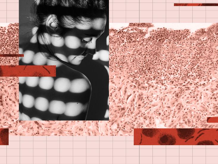
Desmoplastic melanoma is a subtype of cutaneous melanoma, a cancer that begins in melanocytes, the cells that produce skin pigment. The word desmoplastic refers to the dense fibrous tissue that forms around and between the cancer cells. Under a microscope, the tumor may contain spindle-shaped melanoma cells woven through scar-like connective tissue.
It represents only a small fraction of melanomas and is diagnosed most often in older adults. Common locations include the scalp, face, and neck, although it can occur on the trunk or limbs. Many tumors contain little visible pigment, so “watch for a black mole” is not enough.
Pathologists often classify it as pure or mixed desmoplastic melanoma. In pure tumors, most of the invasive cancer has the fibrous pattern. Mixed tumors also contain a more conventional melanoma component. This distinction matters because mixed tumors generally have a greater likelihood of involving nearby lymph nodes, while pure tumors may pose a particularly important local-recurrence problem.
Symptoms: What does desmoplastic melanoma look and feel like?
The lesion is often firm, raised, and skin-colored, pink, red, tan, or lightly pigmented. It may resemble a scar even when no injury occurred. Some tumors form a thick plaque beneath the skin; others appear as a slowly enlarging nodule.
Possible warning signs
- A new firm bump or thickened patch on sun-damaged skin
- A scar-like area without a clear history of injury or surgery
- A pale, pink, reddish, tan, or occasionally dark growth
- A lesion that enlarges, hardens, changes texture, or returns after removal
- Bleeding, crusting, ulceration, or a sore that does not heal
- Itching, tenderness, pain, tingling, burning, numbness, or weakness nearby
The ABCDE checklistAsymmetry, Border irregularity, Color variation, Diameter, and Evolutionstill helps when pigment is present. However, desmoplastic melanoma may ignore several of those rules. Evolution, meaning persistent change over time, is often the best clue. A growing, unexplained, firm lesion deserves evaluation even if it is perfectly beige.
Why it is easily mistaken for something harmless
Desmoplastic melanoma can resemble a scar, cyst, dermatofibroma, basal cell carcinoma, squamous cell carcinoma, or another spindle-cell tumor. Even microscopic diagnosis can be difficult. Dermatopathologists may use the tissue pattern and immunohistochemical markers such as SOX10 or S100 to support the diagnosis and separate it from look-alikes.
Causes and risk factors
There is rarely one identifiable cause in an individual patient. Desmoplastic melanoma develops when genetic damage allows melanocytes to grow and survive abnormally. Long-term ultraviolet radiation is strongly associated with this subtype, which helps explain its preference for chronically sun-exposed skin and its often-high burden of DNA mutations.
Factors associated with higher risk
- Years of cumulative sun exposure
- Older age
- Fair skin, light eyes, freckles, or a tendency to burn
- A personal or family history of melanoma
- A weakened immune system
- Extensive sun-related skin damage
Risk is not destiny. People with darker skin can develop melanoma, and some patients have no obvious risk factors. A diagnosis is also not proof that someone “failed” at sun protection; cancer usually reflects accumulated biological changes, not one forgotten sunscreen application.
How desmoplastic melanoma is diagnosed
Diagnosis begins with a skin examination and history. A dermatologist may ask when the lesion appeared, how it has changed, and whether it causes pain, itching, or altered sensation. Dermoscopy can help assess surface features, but a biopsy is required to confirm melanoma.
Biopsy and pathology review
When practical, the clinician removes the suspicious lesion with a narrow margin for examination. Larger or difficult lesions may require another biopsy approach. Because the cancer can extend deeper or wider than it appears, obtaining enough tissue is important. Expert dermatopathology review can be valuable when the findings are unusual or uncertain.
The pathology report may describe Breslow thickness, ulceration, margins, mitotic activity, the pure or mixed pattern, and neurotropismtumor growth around or within nerves. Neurotropism can explain pain, tingling, or numbness and may raise concern about recurrence near the original site.
Staging and lymph-node evaluation
After diagnosis, the team determines the stage through examination and, when appropriate, sentinel lymph node biopsy or imaging. The role of sentinel node biopsy is individualized. Pure desmoplastic melanoma tends to have a lower rate of lymph-node involvement than mixed disease, so tumor thickness, ulceration, location, overall health, and whether the result would change treatment all belong in the discussion.
Desmoplastic melanoma treatment
Treatment depends on stage, location, nerve involvement, surgical margins, overall health, and whether the cancer can be removed. A multidisciplinary team may include dermatology, surgical oncology, head-and-neck surgery, medical oncology, radiation oncology, plastic surgery, and dermatopathology. Rare tumors are not the ideal setting for medical solo karaoke.
Wide local excision
Surgery is the main treatment for localized disease. The surgeon removes the tumor with a margin of normal-appearing tissue to reduce the chance that microscopic cells remain. Tumors on the face, scalp, or neck may require a graft, flap, or other reconstruction. If cancer is present at the specimen edge, additional surgery may be recommended when feasible.
Sentinel lymph node biopsy
This staging procedure removes the first lymph node or nodes most likely to receive drainage from the tumor area. It is not the same as removing all regional lymph nodes. Clinicians often evaluate mixed tumors similarly to other invasive melanomas of comparable thickness. For pure tumors, the lower average nodal risk makes shared decision-making especially important.
Radiation therapy
Radiation is not routine for everyone, but it may be considered when local-recurrence risk is highfor example, extensive nerve involvement, recurrent disease, close or positive margins that cannot be improved surgically, or a difficult head-and-neck tumor. It may also help control symptoms or disease that cannot be completely removed.
Immunotherapy
Checkpoint inhibitors help the immune system attack melanoma cells. PD-1 drugs such as pembrolizumab and nivolumab are used in selected patients with high-risk, unresectable, or metastatic melanoma. Depending on the stage, immunotherapy may be given after surgery to reduce recurrence risk or used as treatment for advanced disease.
Desmoplastic melanoma appears unusually sensitive to PD-1 blockade in clinical research. A small phase 2 study of unresectable disease reported responses in 24 of 27 patients treated with pembrolizumab. A separate 2026 phase 2 report in resectable disease found a complete pathological response in 20 of 28 evaluable patients after preoperative pembrolizumab. These are encouraging results from limited cohorts, not a guarantee of benefit or a universal treatment formula. Timing, side effects, autoimmune conditions, transplant history, and current guidelines must be considered.
Targeted therapy and clinical trials
Advanced tumors are often tested for actionable mutations. If a BRAF V600 mutation is present, combined BRAF and MEK inhibitors may be an option. Not every desmoplastic melanoma has a targetable alteration, so clinical trials can be important for recurrent, unresectable, or metastatic disease.
Prognosis, recurrence, and follow-up
Prognosis cannot be predicted from the subtype name alone. Stage, Breslow thickness, ulceration, margins, neurotropism, lymph-node status, distant spread, and general health all matter. Desmoplastic melanoma may grow deeply before recognition and has a notable tendency to recur locally or regionally.
Follow-up commonly includes regular skin examinations, evaluation of the surgical area and lymph nodes, and imaging when indicated. People who have had melanoma also face an increased risk of another primary melanoma, making long-term surveillance part of treatment rather than optional paperwork.
Prevention and early detection
No plan eliminates all risk, but reducing ultraviolet exposure helps prevent melanoma and other skin cancers. Use broad-spectrum, water-resistant SPF 30 or higher, wear protective clothing and a wide-brimmed hat, seek shade, avoid indoor tanning, and reapply sunscreen after swimming, sweating, or about every two hours outdoors.
Monthly self-checks can help reveal change. Examine the scalp, ears, neck, back, limbs, hands, feet, nails, and other easy-to-ignore areas. Photograph difficult spots for comparison. See a dermatologist for a new, growing, firm, scar-like, bleeding, painful, tingling, or numb lesionespecially on chronically sun-damaged skin.
Frequently asked questions
Is desmoplastic melanoma always dark?
No. Many tumors are amelanotic, meaning they contain little visible pigment. They may be flesh-colored, pink, red, tan, or scar-like.
Is it more aggressive than ordinary melanoma?
It behaves differently rather than fitting a simple “more” or “less” aggressive label. It can invade deeply and recur locally. Pure tumors may be less likely than mixed tumors to spread to lymph nodes, but stage remains central to prognosis.
Can a scar turn into melanoma?
Most scars do not become melanoma. The issue is that desmoplastic melanoma can imitate a scar. A scar-like area that appears without an injury, grows, hardens, or causes unusual sensations should be examined.
Does a biopsy make melanoma spread?
No credible evidence shows that a properly performed skin biopsy causes melanoma to spread. Biopsy is the standard method for establishing the diagnosis and planning care.
The patient experience: What the journey may feel like
The experience often begins with uncertainty rather than alarm. A person may notice a firm patch near the temple, a pale bump on the scalp, or a thick spot on the neck. It may have seemed harmless for months. A clinician might reasonably think first of a cyst or scar because common conditions are common. When the biopsy unexpectedly says “desmoplastic melanoma,” the emotional shift can be abrupt: yesterday it was an annoying bump; today it has a long medical name and an oncology appointment.
Waiting for pathology details can be one of the hardest stages. Terms such as Breslow depth, neurotropism, margins, pure, and mixed may arrive like guests who forgot to introduce themselves. Bringing a trusted person to appointments, writing questions in advance, and requesting a pathology copy can make the information manageable. Helpful questions include: Are the margins clear? Is the tumor pure or mixed? Was nerve involvement found? Would a sentinel node biopsy change treatment? Should the slides be reviewed by a specialist dermatopathologist?
Surgery may be more involved than expected, particularly on the head or neck. Removing an adequate margin can leave a wound requiring a skin graft, flap, or staged reconstruction. The first look at the surgical site may be emotionally difficult even when healing is normal. Swelling, numbness, tightness, and temporary appearance changes may improve gradually. Clear wound-care instructions, realistic healing photographs, and early contact with the surgical team when something seems wrong can reduce anxiety.
If radiation or immunotherapy is recommended, the experience shifts from one procedure to an ongoing schedule. Radiation may cause localized skin irritation, fatigue, or hair loss in the treated area. Checkpoint inhibitors may be easy for some people and demanding for others. Because they activate immune responses, they can inflame organs including the skin, bowel, liver, lungs, thyroid, and other glands. New diarrhea, shortness of breath, severe rash, yellowing skin, unusual weakness, or persistent headache should be reported promptly rather than saved for the next routine visit.
After treatment, recurrence anxiety may become the quiet challenge. Before follow-up, every twinge can feel suspicious. A practical surveillance routine helps: keep appointments, learn how the surgical area normally feels, photograph the skin, and ask which symptoms require an urgent call. Counseling, melanoma support groups, and honest family conversations can help patients live with uncertainty without letting it run the household.
This is a composite description of experiences reported across melanoma care, not one patient’s story. Some people need surgery alone; others need several specialties and months of treatment. The most useful approach is to focus on the next verified step: expert pathology, accurate staging, a tailored plan, and reliable follow-up.
Conclusion
Desmoplastic melanoma is rare, often nonpigmented, and easy to confuse with a scar or benign growth. Warning signs include a new or changing firm patch, thickening, nodule, unexplained scar-like lesion, bleeding, pain, tingling, or numbness. Diagnosis requires biopsy and careful pathology review. Treatment usually begins with wide local excision, while lymph-node evaluation, radiation, immunotherapy, targeted therapy, or a clinical trial may be appropriate depending on subtype and stage.
When a spot keeps changing or a “scar” appears without a good story, let a dermatologist be the detective.