A heart MRI, also called a cardiac MRI or cardiovascular magnetic resonance imaging, is a detailed imaging test that helps doctors see the heart in action without using X-rays. Think of it as a high-resolution movie camera for your heartminus the popcorn, unfortunately. It uses a strong magnet, radio waves, and computer technology to create clear pictures of the heart muscle, chambers, valves, major blood vessels, and blood flow.
For many people, the phrase “heart test” immediately brings to mind treadmills, sticky electrodes, or someone saying, “Hold still,” while you wonder whether your left sock contains forbidden metal. A heart MRI can sound intimidating, but the test is noninvasive, usually painless, and often extremely useful when other tests do not provide enough detail. Doctors may order it to evaluate heart disease, cardiomyopathy, myocarditis, congenital heart defects, scar tissue after a heart attack, blood flow problems, or unexplained symptoms such as chest pain or shortness of breath.
This guide explains the purpose of a heart MRI, how to prepare, what happens during the procedure, what the results may show, and what real patients often wish they had known before sliding into the MRI scanner like a very cautious burrito.
What Is a Heart MRI?
A heart MRI is a medical imaging test designed specifically to examine the heart and nearby blood vessels. Unlike a standard chest X-ray or CT scan, MRI does not use ionizing radiation. Instead, it uses a powerful magnetic field and radiofrequency pulses to produce detailed images of soft tissues. Because the heart is constantly moving, cardiac MRI uses special techniques to capture motion, measure pumping function, and show blood flow through the heart.
During the scan, the MRI system collects many image “slices.” These slices can be viewed separately or reconstructed into a detailed picture of the heart. Some scans show the heart beating in real time. Others look for inflammation, swelling, scar tissue, or areas with poor blood supply. If contrast is used, a gadolinium-based contrast agent may be injected into a vein to help highlight certain tissues and improve the visibility of blood vessels or damaged heart muscle.
Why Doctors Order a Heart MRI
The main purpose of a heart MRI is to give doctors a deeper look at heart structure and function. It can answer questions that are difficult to solve with a physical exam, blood test, ECG, echocardiogram, or stress test alone. A cardiac MRI is especially valuable because it can show both anatomy and function in one exam.
To Measure Heart Function
One of the most common uses of a heart MRI is measuring how well the heart pumps. The scan can calculate chamber size, wall thickness, stroke volume, and ejection fraction. Ejection fraction is the percentage of blood pumped out of the left ventricle with each heartbeat. This measurement helps doctors diagnose and monitor heart failure, cardiomyopathy, and other conditions that affect pumping strength.
To Evaluate Heart Muscle Damage
Heart MRI can help identify areas of heart muscle that have been injured by a heart attack, inflammation, infection, or reduced blood flow. With contrast imaging, doctors can often distinguish healthy heart muscle from scarred or damaged tissue. This is important when deciding whether a patient may benefit from medications, procedures, surgery, or additional monitoring.
To Diagnose Cardiomyopathy
Cardiomyopathy refers to diseases of the heart muscle. Some forms make the heart enlarged and weak, while others make the muscle thick, stiff, or abnormal in texture. A heart MRI can help identify patterns seen in hypertrophic cardiomyopathy, dilated cardiomyopathy, restrictive cardiomyopathy, arrhythmogenic right ventricular cardiomyopathy, and infiltrative diseases such as cardiac amyloidosis or sarcoidosis.
To Check for Myocarditis
Myocarditis is inflammation of the heart muscle. It may occur after viral infections, autoimmune conditions, or other triggers. Heart MRI is often used because it can detect swelling, inflammation, scarring, and changes in heart function. In some cases, it helps doctors decide whether symptoms are likely due to myocarditis or another heart condition.
To Examine the Pericardium
The pericardium is the thin sac surrounding the heart. A cardiac MRI can help evaluate pericarditis, pericardial thickening, pericardial fluid, constrictive pericarditis, and other pericardial problems. This matters because pericardial disease can sometimes mimic other heart or lung conditions.
To Assess Congenital Heart Disease
For children and adults born with heart defects, cardiac MRI can provide detailed information about heart chambers, blood vessels, shunts, repaired defects, and long-term changes after surgery. It is often used for follow-up because it can measure blood flow and anatomy without radiation exposure.
To Study Blood Flow and Stress Perfusion
A stress cardiac MRI may be ordered to evaluate how blood flows to the heart muscle under stress. Instead of running on a treadmill, many patients receive a medication that temporarily makes the heart respond as if it were exercising. Images are taken before, during, and after stress to look for areas that may not be receiving enough blood.
Heart MRI vs. Other Heart Tests
A heart MRI does not replace every other cardiac test. Instead, it fills a specific role. An echocardiogram is often faster and more widely available, making it a common first test for heart structure and function. A CT scan may be better for imaging coronary artery calcium or certain coronary artery anatomy. A nuclear stress test can assess blood flow using radioactive tracers. Cardiac catheterization can directly measure pressures and treat blocked arteries.
Heart MRI stands out because it offers excellent soft-tissue detail, accurate measurements of heart size and pumping function, and the ability to characterize heart muscle tissue. In plain English: it does not just ask, “Is the heart shaped correctly?” It asks, “How is the heart moving, where is the damage, and what kind of damage might it be?” Very nosybut medically useful.
How to Prepare for a Heart MRI
Preparation depends on the type of heart MRI being performed. A standard cardiac MRI may require very little preparation. A stress cardiac MRI, contrast-enhanced MRI, or sedated MRI may involve extra instructions. Always follow the directions from your imaging center, because protocols vary.
Tell the Team About Implants and Metal
Before the scan, you will complete an MRI safety screening form. Be honest and detailed. The MRI magnet is powerful, so the team needs to know if you have a pacemaker, implantable cardioverter-defibrillator, artificial heart valve, aneurysm clip, cochlear implant, insulin pump, neurostimulator, metal fragments, surgical hardware, stents, implanted ports, or any other device.
Many modern devices are MRI-conditional, meaning they may be safe under specific settings. However, the radiology team must verify this before the scan. Do not guess. “I think it is fine” is not the same as “the device documentation confirms MRI safety.” Your heart deserves paperwork, not vibes.
Remove Jewelry and Metal Objects
You will usually be asked to remove jewelry, watches, hairpins, hearing aids, dentures, glasses, piercings, credit cards, keys, and clothing with metal zippers or snaps. Some cosmetics, underwire bras, and clothing fibers may contain metal, so many centers provide a hospital gown. This is not a fashion moment. It is a safety moment wearing a back-opening curtain.
Ask About Food, Drink, and Medications
For many MRI exams, patients can eat, drink, and take regular medications as usual. However, some cardiac MRI exams require fasting for several hours. If you are having a stress cardiac MRI, you may be told to avoid caffeine for a specific period before the test because caffeine can interfere with stress medications. This includes coffee, tea, energy drinks, chocolate, some sodas, and certain medications that contain caffeine.
Do not stop prescription medications unless your healthcare provider tells you to. If you take medicines for diabetes, blood pressure, heart rhythm, or chest pain, ask for specific instructions before the appointment.
Discuss Kidney Disease and Contrast
Some heart MRI scans use gadolinium-based contrast. In most patients, MRI contrast is well tolerated, but people with significant kidney disease may need special evaluation. Your doctor may order a kidney function blood test before the scan. Tell the team if you have chronic kidney disease, are on dialysis, have had a kidney transplant, or have had a previous reaction to MRI contrast.
Mention Pregnancy or Breastfeeding
If you are pregnant, might be pregnant, or are breastfeeding, tell your healthcare provider and the radiology team. MRI without contrast may be used during pregnancy when medically necessary, but contrast use is considered carefully. Your care team will balance the benefits and risks based on your situation.
Plan for Claustrophobia
The MRI scanner is a tube-shaped machine, and some people feel anxious in tight spaces. If you have claustrophobia, tell your doctor before the appointment. You may be offered relaxation strategies, music, a mirror, extra communication with the technologist, or medication to help you relax. If you receive a sedative, you will likely need someone to drive you home.
What Happens During the Heart MRI Procedure?
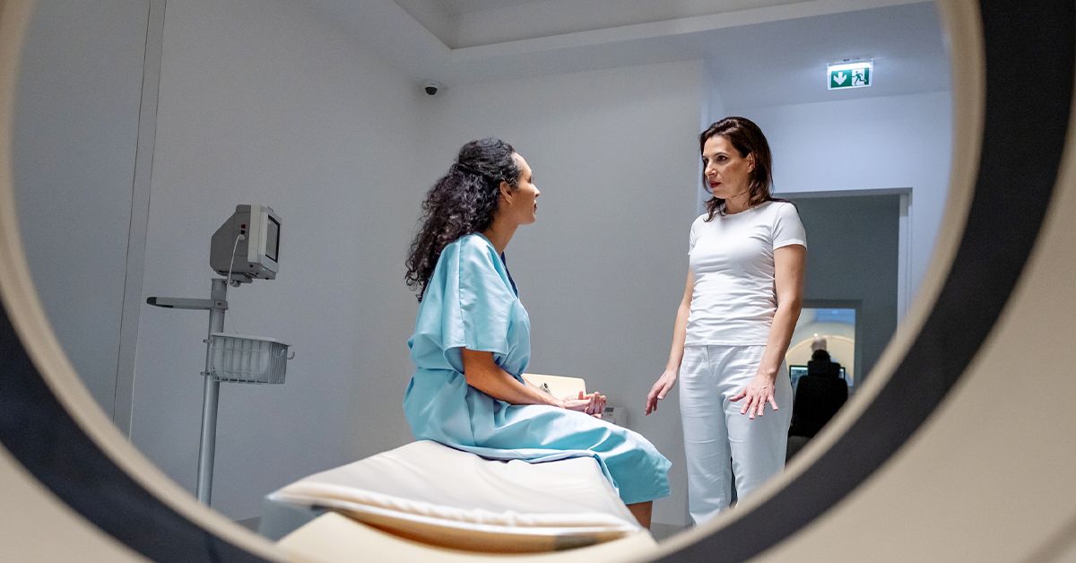
On the day of the test, you will check in, complete safety forms, change if needed, and review your medical history with the technologist or nurse. The team may place ECG leads on your chest to synchronize the images with your heartbeat. This helps the scanner capture images at the right points in the cardiac cycle.
Step 1: Positioning on the Table
You will lie on a padded table that slides into the MRI scanner. A special receiver coil may be placed over your chest to improve image quality. The technologist will make sure you are positioned comfortably, because movement can blur the pictures. You may be given earplugs or headphones because MRI machines make loud knocking, tapping, and thumping sounds. The scanner is not broken; it is just dramatic.
Step 2: Breath-Holding Instructions
For many cardiac MRI sequences, you will be asked to hold your breath for short periods. This reduces motion from breathing and helps create sharper images. A typical instruction may sound like: “Breathe in, breathe out, hold your breath.” Each breath-hold may last several seconds. If you cannot hold your breath well, tell the technologist. Modern MRI techniques can often be adjusted.
Step 3: Imaging Sequences
The scan usually includes several imaging sequences. Cine images show the heart beating. Black-blood images may show anatomy. Flow imaging may measure blood movement through valves or vessels. Tissue characterization sequences may look for swelling, fat, iron, scar, or inflammation. If contrast is used, images may be taken shortly after injection and again later to look for delayed enhancement.
Step 4: Contrast or Stress Medication
If your exam requires contrast, a small IV will be placed in your arm or hand. You may feel a cool sensation during injection. If you are having a stress cardiac MRI, you may receive medication that temporarily changes blood flow in the heart. Some people feel flushed, short of breath, warm, lightheaded, or notice chest pressure during stress medication. These feelings usually pass quickly, and the team monitors you throughout the test.
Step 5: Finishing the Scan
A heart MRI can take around 30 to 90 minutes, depending on the protocol. Some complex exams may take longer. After the scan, the table slides out, the IV is removed if one was placed, and you can usually return to normal activities. If you received a sedative, you should not drive, drink alcohol, or make important decisions until it wears off. That includes online shopping for “medical recovery snacks” in bulk.
Is a Heart MRI Safe?
For most people, a heart MRI is considered safe and noninvasive. It does not use radiation. The main safety concern is the strong magnet, which can affect certain implants, devices, and metal objects. That is why MRI screening is so important.
Gadolinium contrast is generally used only when it adds diagnostic value. Allergic reactions are uncommon, but they can happen. Patients with severe kidney disease require special consideration because some gadolinium agents have been linked to a rare condition called nephrogenic systemic fibrosis. Modern screening and contrast selection have greatly reduced this risk, but it is still important to disclose kidney problems.
What Can Heart MRI Results Show?
After the scan, a radiologist or cardiologist trained in cardiac imaging reviews the images. Your ordering provider will discuss the results with you. The report may include measurements of heart chamber size, wall thickness, pumping function, valve function, blood flow, scar tissue, inflammation, pericardial findings, masses, congenital anatomy, or blood vessel abnormalities.
Results may help confirm a diagnosis, rule out serious conditions, guide treatment, monitor disease progression, or evaluate whether a previous treatment is working. For example, if a patient has unexplained shortness of breath, a heart MRI may show whether the problem is related to weak pumping, thickened heart muscle, inflammation, or scarring. If someone has had a heart attack, MRI may help determine how much heart muscle is damaged and whether some tissue may recover.
Common Questions About Heart MRI
Does a heart MRI hurt?
No, the scan itself should not hurt. You may feel discomfort from lying still, holding your breath, or having an IV placed. The machine is loud, but ear protection helps.
Can I move during the scan?
Small movements can blur the images, so you should stay as still as possible. If you are uncomfortable, use the call button or tell the technologist between sequences.
Will I be completely inside the scanner?
For a heart MRI, your chest must be centered in the scanner, so much of your body may be inside the tube. Your head may be inside or near the opening depending on your height and scanner design.
Can I drive home afterward?
Most people can drive home after a heart MRI. If you received sedation or certain anxiety medications, arrange a ride home.
How soon will I get results?
Timing varies by facility. Some results are available within a few days, while complex cardiac MRI reports may take longer because measurements and specialized image review require careful analysis.
Experiences Related to Heart MRI: What Patients Often Notice
Many people walk into a heart MRI appointment expecting the worst and leave thinking, “That was weird, but manageable.” The experience is often less painful than imagined, though it can be more mentally demanding than a quick blood test or ECG. The hardest parts are usually lying still, hearing the machine’s loud sounds, and following breath-hold instructions. The scanner may thump, knock, buzz, and hum like it is auditioning for an industrial music festival. Earplugs or headphones make a big difference.
Patients often say the breath-holding is the most surprising part. During a cardiac MRI, the technologist may give repeated instructions to breathe in, breathe out, and hold. These breath-holds help reduce motion so the images are clear. If you miss one, do not panic. The technologist can repeat a sequence if needed. The goal is not Olympic-level breath control. The goal is cooperation, communication, and doing your best.
Another common experience is feeling nervous before contrast injection. Most people feel little more than the IV placement and perhaps a cool sensation. If stress medication is used, the experience can be more noticeable. Some patients feel warmth, flushing, shortness of breath, or pressure in the chest. These sensations can be unsettling, but they are expected with certain stress agents and usually fade quickly. The medical team monitors your heart rhythm, symptoms, and overall response during the test.
Claustrophobia is also common. Even people who are normally calm may feel uneasy once the table moves into the scanner. Practical tricks can help: close your eyes before entering the scanner, focus on breathing slowly, ask whether music is available, and remind yourself that you can communicate with the technologist. Some people mentally divide the scan into small segments: “Just this breath-hold,” then “just this next sound,” then “just the next few minutes.” It is not glamorous, but it works.
People who prepare well tend to have smoother appointments. Wearing easy-to-change clothing, leaving jewelry at home, avoiding caffeine if instructed, bringing implant information, and arriving early can prevent delays. If you have a pacemaker, defibrillator, valve replacement, stent, surgical clip, or any implanted device, bring the device card if you have it. This can save time and reduce uncertainty.
After the scan, many patients feel relieved more than anything else. The procedure usually does not require recovery unless sedation was used. You can typically eat, drink, work, and return to regular activities. If contrast was given, drinking water afterward may be suggested unless you have fluid restrictions. The waiting period for results can be the most emotionally difficult part. It is normal to feel anxious, but remember that the scan is a tool for clarity. Whether it confirms a problem, rules one out, or helps guide treatment, the purpose is to give your healthcare team better information.
The best mindset for a heart MRI is simple: prepare honestly, communicate clearly, and remember that the machine may be loud, but it is on your side. It is there to take pictures, not judge your cholesterol history or your relationship with drive-thru fries.
Conclusion
A heart MRI is one of the most detailed noninvasive tests available for evaluating the heart. It can measure pumping function, examine heart muscle, identify scar tissue, detect inflammation, assess congenital heart problems, evaluate the pericardium, and help guide treatment decisions. Preparation usually focuses on safety screening, metal removal, medication instructions, contrast considerations, and comfort planning.
Although the scan may feel intimidating, most people tolerate it well. The key is knowing what to expect: a loud machine, a padded table, breath-hold instructions, possible contrast or stress medication, and a team monitoring you throughout the exam. A heart MRI may not be anyone’s idea of a spa day, but when it comes to understanding the heart, it is a powerful diagnostic tool with a very important job.
Note: This article is for educational purposes only and should not replace medical advice from a qualified healthcare professional. Always follow the instructions provided by your doctor or imaging center.