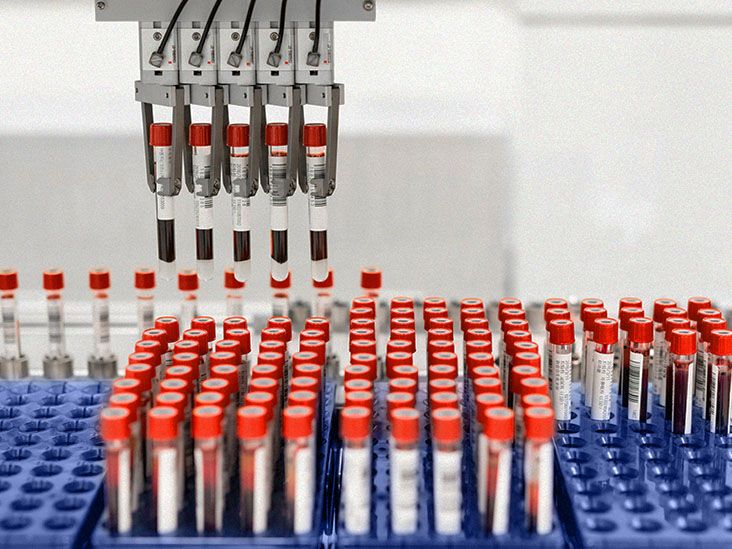
Small cell lung cancer tumor markers can sound like secret codes from a medical spy movie: NSE, ProGRP, CD56, Ki-67, DLL3, LDH. The names are tiny; the implications are not. These markers help doctors understand whether a lung tumor has the “signature” of small cell lung cancer (SCLC), how active the cancer may be, whether treatment is working, and whether a clinical trial or newer therapy might be worth discussing.
Here is the most important thing to know right away: tumor markers do not diagnose small cell lung cancer by themselves. They are clues, not courtroom verdicts. A biopsy, imaging, pathology review, and clinical context remain the main tools for diagnosis and staging. Still, tumor markers can be incredibly useful. Think of them as the sticky notes your cancer care team uses to organize a very complicated case file.
This guide explains the major types of SCLC tumor markers, typical reference ranges, how results are interpreted, and what patients and caregivers often experience while trying to make sense of these tests.
What Are Tumor Markers in Small Cell Lung Cancer?
Tumor markers are measurable substances or features linked to cancer cells. In SCLC, they may be proteins seen under a microscope, substances measured in blood, genetic changes, or newer treatment-related targets. Some markers help pathologists confirm that a tumor is neuroendocrine in nature. Others help oncologists estimate tumor burden, monitor response to treatment, or evaluate whether a patient may qualify for a specific therapy or clinical trial.
Small cell lung cancer is a fast-growing, high-grade neuroendocrine cancer. It accounts for a smaller share of lung cancers than non-small cell lung cancer, but it tends to spread earlier and act more aggressively. That is why doctors usually move quickly: biopsy, scan, stage, treat. SCLC does not like waiting rooms either.
Why Tumor Markers Matter in SCLC
SCLC tumor markers may help in several ways:
- Diagnosis: Immunohistochemistry markers can support a diagnosis of SCLC when tissue is examined by a pathologist.
- Disease monitoring: Blood markers such as neuron-specific enolase (NSE) and pro-gastrin-releasing peptide (ProGRP) may rise or fall with tumor activity in some patients.
- Prognosis: Some markers, including high LDH or very elevated NSE, may suggest a larger disease burden or more aggressive behavior.
- Treatment planning: Emerging markers such as DLL3, SLFN11, and molecular subtypes are becoming more relevant as SCLC research advances.
- Clinical trials: Biomarker testing may identify trial options, especially when standard treatments stop working.
But there is a catch: many tumor markers are not specific to SCLC. A marker may be high because of another cancer, kidney problems, liver injury, inflammation, smoking, blood sample handling, or even plain old lab variation. In other words, tumor markers are helpful, but they are not magic crystal balls wearing white coats.
Main Types of Small Cell Lung Cancer Tumor Markers
1. Immunohistochemistry Markers
Immunohistochemistry, often shortened to IHC, is a lab technique used on biopsy tissue. It uses special stains to detect proteins in tumor cells. For SCLC, IHC markers are especially important because the cancer often has neuroendocrine features.
Common IHC markers for SCLC include:
- Synaptophysin: A neuroendocrine marker often positive in SCLC.
- Chromogranin A: Another neuroendocrine marker, sometimes less sensitive than synaptophysin or CD56.
- CD56: A sensitive neuroendocrine marker, though not perfectly specific.
- INSM1: A newer nuclear marker that can be very useful for identifying neuroendocrine differentiation.
- TTF-1: Often positive in lung-origin SCLC, but not exclusive to SCLC.
- Ki-67: A proliferation marker that shows how rapidly tumor cells are dividing.
In many SCLC tumors, Ki-67 is high, often far above what is seen in slower-growing neuroendocrine tumors such as typical carcinoid tumors. A high Ki-67 index supports the diagnosis of a high-grade tumor, but it does not replace the pathologist’s full review of cell shape, growth pattern, crush artifact, necrosis, and other microscopic features.
2. Serum Tumor Markers
Serum tumor markers are measured in blood. In SCLC, the most discussed serum markers are NSE and ProGRP. Doctors may also follow LDH or CEA in selected cases, though these are less specific.
Blood markers are usually more useful for monitoring trends than for making a diagnosis. A single number can mislead. A pattern over time, matched with scans and symptoms, is much more informative. One blood test is a snapshot; several tests over time are the movie.
3. Molecular and Genomic Markers
Compared with non-small cell lung cancer, SCLC has fewer routine FDA-approved targeted biomarker pathways. However, research has identified common genetic alterations, including loss or inactivation of TP53 and RB1. These changes help explain the biology of SCLC but are not usually “actionable” in the same way that EGFR or ALK alterations may be in non-small cell lung cancer.
Researchers are also studying SCLC subtypes based on transcription factors and immune features, including ASCL1, NEUROD1, POU2F3, and an inflamed subtype sometimes described as SCLC-I. These are not everyday clinic tests for every patient yet, but they are important in research and may shape future treatment decisions.
4. Treatment-Related Emerging Markers
One of the most important emerging targets in SCLC is DLL3, short for delta-like ligand 3. DLL3 is commonly expressed on SCLC cells and has become relevant because newer therapies can target it. Tarlatamab, a DLL3-directed bispecific T-cell engager, is one example of how a tumor marker can move from “interesting lab finding” to “real treatment conversation.”
Other emerging markers include SLFN11, which may relate to sensitivity to DNA-damaging therapies, and immune-related markers such as PD-L1 and tumor mutational burden. Unlike in many non-small cell lung cancers, PD-L1 testing has not been a consistently reliable stand-alone predictor in SCLC. The science is moving, but it has not become a simple “positive means yes, negative means no” situation.
Common SCLC Tumor Markers and Typical Ranges
Reference ranges vary by laboratory, testing method, specimen type, and patient factors. Always use the reference range printed on the actual lab report. The ranges below are general examples, not universal rules.
| Marker | Type | Typical Reference Range or Pattern | How It May Be Used in SCLC |
|---|---|---|---|
| NSE | Blood tumor marker | Often about ≤12.7 to ≤15 ng/mL, depending on lab | May help monitor disease burden and treatment response |
| ProGRP | Blood tumor marker | Often below about 50 to 75 pg/mL, depending on assay | Useful supportive marker for SCLC; may be followed over time |
| CEA | Blood tumor marker | Often 0 to 3 ng/mL in nonsmokers; up to about 5 ng/mL may be seen in smokers | Less specific; may be elevated in some lung cancers and benign conditions |
| LDH | Blood chemistry marker | Common adult ranges are roughly 125 to 225 U/L, depending on lab | May reflect tissue damage, tumor burden, or aggressive disease, but is nonspecific |
| Synaptophysin | IHC tissue marker | Reported as positive or negative staining | Supports neuroendocrine differentiation |
| Chromogranin A | IHC tissue marker | Reported as positive or negative staining | Supports neuroendocrine differentiation, though sensitivity can vary |
| CD56 | IHC tissue marker | Reported as positive or negative staining | Commonly positive in SCLC; sensitive but not fully specific |
| INSM1 | IHC tissue marker | Reported as nuclear staining positive or negative | Helps identify neuroendocrine tumors, including SCLC |
| Ki-67 | IHC proliferation marker | SCLC is usually high; often reported as a percentage | Helps distinguish high-grade SCLC from lower-grade neuroendocrine tumors |
| DLL3 | Tissue/treatment-related marker | Reported by test method; no single universal patient-facing “normal range” | Relevant to DLL3-targeted therapies and clinical trials |
NSE: Neuron-Specific Enolase
NSE is one of the classic serum markers associated with SCLC. It is an enzyme found in nerve and neuroendocrine cells. Since SCLC is a neuroendocrine carcinoma, NSE can be elevated when tumor burden is high.
Typical reference ranges vary. Some labs list normal NSE as less than or equal to about 12.7 ng/mL, while others use cutoffs around 14 or 15 ng/mL. In SCLC, an elevated NSE may support suspicion, but it cannot confirm the diagnosis. NSE can also rise in other neuroendocrine tumors, neurologic injury, hemolysis, and certain noncancer conditions.
A practical example: if a patient’s NSE is 68 ng/mL before treatment and falls to 18 ng/mL after chemotherapy, that trend may fit with treatment response, especially if scans also show tumor shrinkage. But if NSE rises slightly while scans are stable, the doctor may repeat the test, check sample quality, and look at the bigger picture before making treatment changes.
ProGRP: Pro-Gastrin-Releasing Peptide
ProGRP is another important blood marker in SCLC. It is often considered more specific for SCLC than NSE, although it still has limitations. ProGRP may be especially useful when doctors are trying to distinguish SCLC from other lung cancer types or when monitoring response after treatment.
Typical ProGRP cutoffs vary by assay. Many labs consider values below roughly 50 to 75 pg/mL as within the expected range, but the exact number depends on the test platform. Kidney function matters, too. ProGRP can be falsely elevated in people with reduced kidney function, so doctors interpret it carefully.
If ProGRP drops sharply after treatment, that can be encouraging. If it begins rising again after a period of control, it may prompt closer follow-up or repeat imaging. Still, ProGRP should not be treated like a scoreboard that defines the entire game. Symptoms, scans, physical exam, and treatment history all matter.
CEA and LDH: Helpful, But Not SCLC-Specific
CEA, or carcinoembryonic antigen, is better known in colorectal cancer but may be elevated in lung cancers and several benign conditions. Smoking can also raise CEA. Typical levels are often below 3 ng/mL in nonsmokers and may be up to about 5 ng/mL in smokers. In SCLC, CEA is not usually the star of the show, but it may appear in the supporting cast.
LDH, or lactate dehydrogenase, is not a tumor marker in the narrow sense. It is an enzyme released when cells are damaged. LDH can rise from cancer, liver disease, muscle injury, infection, anemia, or many other conditions. In SCLC, high LDH may suggest a larger tumor burden or more aggressive disease, but it is far too nonspecific to interpret alone.
IHC Markers: The Pathology Detective Work
When doctors suspect SCLC, the biopsy tissue usually goes to a pathology lab. Under the microscope, SCLC often appears as small cells with scant cytoplasm, finely granular chromatin, nuclear molding, many mitoses, and areas of necrosis. That description may sound like a villain origin story, but for pathologists, it is useful evidence.
IHC stains then help confirm the diagnosis. A typical SCLC profile may include positivity for synaptophysin, CD56, chromogranin A, INSM1, and TTF-1, with a high Ki-67 proliferation index. However, not every case checks every box. Some tumors stain weakly. Some biopsies are tiny. Some samples are crushed. Pathologists often use a panel of markers rather than relying on one test.
Can Tumor Markers Detect SCLC Early?
At this time, SCLC tumor markers are not recommended as stand-alone screening tests for the general population. Low-dose CT screening is the established tool for eligible high-risk people, such as certain adults with a significant smoking history. Blood markers may someday play a bigger role in early detection, but we are not there yet.
The problem is sensitivity and specificity. A screening test must catch disease early without creating a parade of false alarms. SCLC serum markers can miss early disease and can also be elevated for reasons unrelated to SCLC. That is a tough combo: too many false negatives and false positives. Nobody wants a smoke detector that ignores fire but screams when toast is slightly ambitious.
How Doctors Interpret Tumor Marker Results
Doctors rarely ask, “Is this marker high?” and stop there. They ask better questions:
- Was the blood sample handled correctly?
- Does the marker match the biopsy and imaging findings?
- Is the marker rising, falling, or stable over time?
- Could kidney function, liver injury, smoking, or hemolysis explain the result?
- Are symptoms improving or getting worse?
- Do CT, PET, MRI, or bone scans support the same story?
This is why the “normal range” is only the beginning. A value slightly above normal may be less concerning than a value that doubles repeatedly. A high number that falls with treatment may be useful. A marker that was never elevated at diagnosis may not be useful for monitoring that patient later.
Questions to Ask Your Cancer Care Team
If you or a loved one has SCLC, consider asking:
- Which tumor markers were tested on my biopsy?
- Were neuroendocrine markers positive?
- What was my Ki-67 score, and what does it mean?
- Are NSE or ProGRP useful markers in my case?
- Should my markers be checked during treatment or follow-up?
- Could kidney function, smoking, or sample handling affect my results?
- Are there biomarker-based clinical trials I should consider?
- Is DLL3 testing relevant for my treatment options?
Limitations and Common Misunderstandings
The biggest misunderstanding is thinking that a tumor marker number equals a diagnosis. It does not. A high NSE does not automatically mean SCLC. A normal NSE does not rule it out. A positive CD56 stain supports neuroendocrine differentiation but is not enough on its own. Tumor markers are pieces of a puzzle, and SCLC is not the kind of puzzle you solve with one corner piece and a confident shrug.
Another misunderstanding is assuming that every patient needs every marker. Testing should be guided by the clinical situation. Some markers are routine in pathology. Some are optional. Some are mainly research tools. Some may matter only when considering a clinical trial or later-line therapy.
Conclusion
Small cell lung cancer tumor markers help doctors identify, classify, monitor, and increasingly personalize care for SCLC. The most common tissue markers include synaptophysin, chromogranin A, CD56, INSM1, TTF-1, and Ki-67. Important blood markers include NSE and ProGRP, while CEA and LDH may provide additional context but are less specific. Emerging markers such as DLL3, SLFN11, and molecular subtypes are shaping the future of SCLC treatment research.
The key is interpretation. Tumor marker ranges are lab-dependent, and results must be read alongside biopsy findings, imaging, symptoms, kidney and liver function, treatment history, and overall health. In the right context, these markers can be powerful tools. Used alone, they are more like breadcrumbs: helpful, but not the whole sandwich.
Patient and Caregiver Experiences: What Tumor Marker Testing Can Feel Like
For many patients, the hardest part of tumor marker testing is not the blood draw or the biopsy stain. It is the waiting. A person may hear that their doctor ordered NSE, ProGRP, Ki-67, or additional tissue testing, and suddenly every unfamiliar abbreviation feels like a tiny thundercloud. The lab report arrives with numbers, flags, percentages, and medical phrases that seem written for someone with three degrees and a microscope in the garage.
One common experience is “range anxiety.” A patient sees that a marker is above the reference range and immediately thinks the worst. That reaction is understandable. Cancer turns normal uncertainty into a full-contact sport. But tumor markers are rarely interpreted as isolated numbers. A care team may care more about whether the number is falling after treatment, rising over several tests, or matching what scans show. A single abnormal marker can be scary, but it is not always a turning point.
Caregivers often become the unofficial record keepers. They save lab reports, highlight values, compare dates, and bring notebooks to appointments. This can be helpful, especially when treatment moves quickly. A simple chart with test date, marker value, treatment cycle, scan result, and major symptoms can make conversations with the oncology team clearer. It also helps prevent the classic “I had a question, but it escaped like a raccoon through a dog door” moment during a busy appointment.
Another real-world issue is mixed emotion when markers improve. If NSE or ProGRP drops after treatment, patients may feel hopeful but cautious. SCLC often responds well at first, yet recurrence is common. That emotional push-pull can be exhausting. It is possible to celebrate good results while still asking practical questions about next steps, follow-up scans, maintenance therapy, clinical trials, and symptom management.
Some patients also feel frustrated when markers do not give clear answers. A scan may look stable while a blood marker rises slightly. Or a marker may be normal even when the cancer is active. This does not mean the test was useless. It may mean that the marker is not reliable for that individual, or that the change is too small to act on without more information. In SCLC care, doctors often need several pieces of evidence before changing treatment.
Patients considering newer treatments or trials may hear about DLL3, SLFN11, circulating tumor DNA, or molecular subtypes. This can feel both exciting and overwhelming. The best approach is to ask whether a marker is standard, optional, experimental, or trial-related. Those four categories can make the conversation much less foggy.
Finally, tumor marker testing often teaches patients a broader lesson: cancer care is not just about numbers. Breathing easier, coughing less, eating better, sleeping better, walking farther, and feeling more like oneself are also meaningful signals. Lab values matter, but so does the person attached to the lab values. That person deserves clear explanations, timely answers, and permission to ask the same question twice if the first explanation sounded like alphabet soup.
Note: This article is for educational purposes only and should not replace medical advice, diagnosis, or treatment from an oncology professional. Tumor marker ranges vary by laboratory, and every result should be interpreted by a qualified healthcare provider.