Osteoporosis can weaken bones for years withou clue is a fracture after a small fall or an ordinary movement that should not have caused major damage. Testing can identify low bone density earlier, estimate fracture risk, and help a healthcare professional decide whether monitoring, lifestyle changes, or medication may be appropriate.
The main osteoporosis test is dual-energy X-ray absorptiometry, called DXA or DEXA. A full evaluation may also include a medical history, physical examination, fracture-risk calculation, laboratory tests, and spine imaging. Here is what each test does, how to prepare, and what the results actually mean.
What Is an Osteoporosis Test?
An osteoporosis evaluation looks at bone mineral density, or BMD, plus the other factors that influence whether a bone is likely to break. BMD reflects how much mineral is packed into a measured area of bone. Denser bone is generally stronger, but age, falls, previous fractures, medications, and medical conditions also matter.
Central DXA is the standard measurement because it usually checks the hip and lumbar spine, two important fracture sites. It can detect low bone density, support an osteoporosis diagnosis, estimate fracture risk, and monitor treatment. The test is noninvasive, painless, and uses very low radiation. Should Have Osteoporosis Screening?
Women age 65 and older
The U.S. Preventive Services Task Force recommends screening women age 65 and older. It also recommends screening postmenopausal women younger than 65 when a clinical risk assessment shows increased fracture risk.
Men and younger adults
The USPSTF says evidence is insufficient to recommend routine population screening for men. However, other professional guidance supports DXA for many men age 70 and older and for younger men with risk factors. Testing may also be appropriate at any adult age when a disease, medication, or fracture suggests abnormal bone loss. mon reasons for earlier testing
- A fracture after a minor fall or ordinary activity, especially after age 50
- Long-term use of glucocorticoids such as prednisone
- Very low body weight, an eating disorder, early menopause, low estrogen, or low testosterone
- A parent with osteoporosis or a hip fracture
- Smoking, heavy alcohol use, or repeated falls
- Rheumatoid arthritis, celiac disease, inflammatory bowel disease, thyroid disease, kidney disease, or another bone-affecting condition
- Height loss, stooped posture, or unexplained back pain that could signal a spinal fracture
Bone Density Scan: What Happens During a DXA Test?
A DXA scanner uses two low-dose X-ray beams to separate bone from soft tissue and calculate density. The hip and spine are usually measured. The forearm may be used when the hip or spine cannot be interpreted accurately because of severe arthritis, surgery, hardware, or another problem.
How to prepare
- Avoid calcium supplements for about 24 hours unless the imaging center gives different instructions.
- Wear loose clothing without metal zippers, buckles, snaps, or underwire.
- Remove jewelry, keys, coins, and other metal objects.
- Tell the staff if you are pregnant or might be pregnant.
- Report a recent barium exam, contrast-enhanced CT, or nuclear medicine study. You may need to postpone DXA, sometimes for roughly 10 to 14 days.
Most people can eat, drink, and take their usual medicines normally. Always follow the specific instructions from the facility performing the scan. ing the appointment
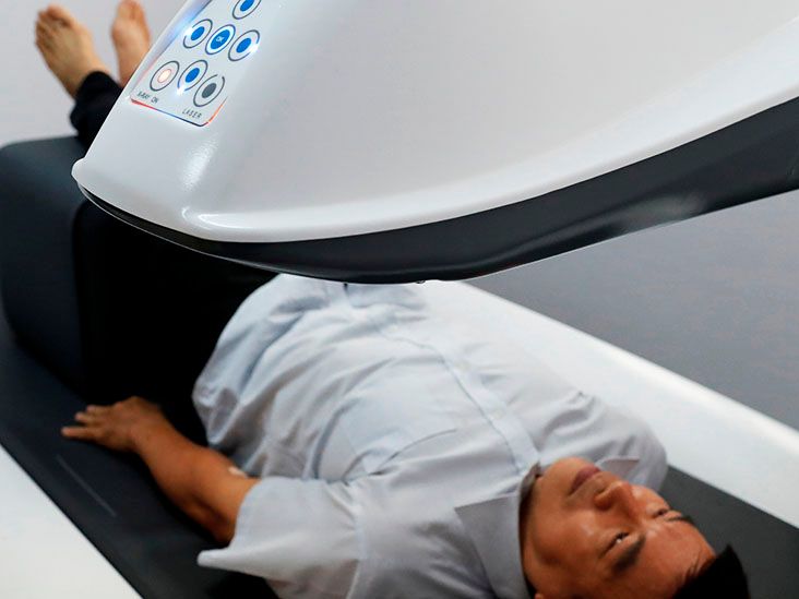
You lie on your back on an open, padded table. A technologist may place a foam block under your legs for the spine scan or position one foot to measure the hip. A scanning arm passes above the body without touching it. There is no tunnel, injection, or sedation.
You must stay still and may briefly hold your breath. The exam often takes about 10 to 30 minutes. It should not hurt, although people with hip or back pain may need extra support. Afterward, you can return to normal activities immediately. erstanding Bone Density Results
T-score
For postmenopausal women and men age 50 or older, results are commonly reported as a T-score. This compares your BMD with the average peak density of a healthy young adult.
| T-score | Usual interpretation |
|---|---|
| -1.0 or higher | Normal bone density |
| Below -1.0 and above -2.5 | Low bone mass, often called osteopenia |
| -2.5 or lower | Bone density in the osteoporosis range |
A T-score is not a complete verdict. A hip or vertebral fragility fracture can support an osteoporosis diagnosis even when the score is better than -2.5. Treatment decisions also consider age, falls, medication exposure, and overall fracture risk. core
A Z-score compares BMD with people of similar age and sex. It is emphasized for premenopausal women, men younger than 50, and children. A Z-score of -2.0 or lower is considered below the expected range for age and may prompt testing for an underlying disease, nutritional problem, hormone disorder, or medication effect. hip and spine results may differ
Arthritis, scoliosis, old compression fractures, calcification, or surgical hardware can distort a spine result. An experienced reader reviews the images and may exclude unreliable vertebrae rather than trusting the boldest number on the page. er Tests Used to Evaluate Osteoporosis
Medical history and physical examination
Your clinician may review fractures, falls, family history, diet, exercise, smoking, alcohol, menopause, menstrual history, testosterone symptoms, digestive problems, kidney stones, and medicines. Height loss or a curved upper back may suggest vertebral compression fractures.
Blood and urine tests
DXA can show low density, but it cannot explain the cause. After osteoporosis is diagnosedor when bone density is unexpectedly lowtesting may include a complete blood count, metabolic panel, 25-hydroxyvitamin D, parathyroid hormone, phosphate, and a 24-hour urine calcium measurement. Depending on the history, a clinician may add thyroid, testosterone, celiac, liver, kidney, or blood-disorder testing. X fracture-risk assessment
FRAX estimates the 10-year probability of hip fracture and major osteoporotic fracture. It combines age, sex, height, weight, prior fracture, parental hip fracture, smoking, glucocorticoid use, rheumatoid arthritis, alcohol exposure, and sometimes femoral-neck BMD. It is especially useful when DXA shows osteopenia rather than osteoporosis. tebral fracture assessment and X-rays
A vertebral fracture assessment, or VFA, is a low-radiation side-view image that may be performed on the DXA machine. It can reveal silent compression fractures. Standard spine X-rays may be ordered for height loss, new back pain, or posture changes. Regular X-rays can show fractures but cannot reliably measure early bone loss. ipheral scans, heel ultrasound, and CT
Portable devices may measure the wrist, finger, or heel. Heel ultrasound uses no radiation and can identify people who need further evaluation, but it usually does not replace central DXA for diagnosis or treatment monitoring. Quantitative CT can provide three-dimensional density information in selected cases, although it is less widely used and generally involves more radiation. e turnover markers and specialized tests
Blood or urine markers such as CTX, NTX, or P1NP reflect bone breakdown or formation. They do not diagnose osteoporosis by themselves but may help monitor biological response to treatment. Bone biopsy is rarely needed. A nuclear bone scan is a different test used to investigate cancer, infection, inflammation, or certain injuriesnot routine osteoporosis. t Can Affect DXA Accuracy?
Severe arthritis, scoliosis, compression fractures, vascular calcification, obesity beyond equipment limits, joint replacements, and metal hardware can interfere with measurement. For follow-up scans, using the same facility and machine can improve comparison because manufacturers and software may calculate results differently.
A small numerical change is not always a true biological change. The report should indicate whether the difference exceeds the facility’s expected measurement error, often called the least significant change. t Happens After an Osteoporosis Test?
The next step depends on the lowest reliable score, fracture history, FRAX estimate, fall risk, medical conditions, kidney function, and treatment preferences. Recommendations may include resistance and weight-bearing exercise, balance training, adequate calcium and protein intake, correction of vitamin D deficiency, smoking cessation, fall prevention, or prescription medication.
Repeat testing is individualized. Someone with normal density and few risks may wait several years. A person starting treatment, taking a bone-weakening drug, or sitting near a treatment threshold may be reassessed sooner. Medicare commonly covers eligible bone mass measurements once every 24 months and more often when medically necessary, but coverage and clinical timing are not identical. t the Osteoporosis Testing Experience Often Feels Like
Many people arrive for their first bone density scan expecting a large hospital machine, a needle, or at least a dramatic mechanical hum worthy of a science-fiction movie. The real appointment is usually much less theatrical. Check-in may take longer than the scan. After confirming pregnancy status, recent imaging, medicines, and supplements, the technologist may ask you to remove metal objects or change into a gown.
The DXA room feels more like a standard imaging room than an MRI suite. The table is open on every side, and the scanner’s arm moves above you without touching you. For the spine measurement, your lower legs may rest on a padded block. For the hip measurement, a positioning device may turn the leg inward. That angle can feel mildly awkward, especially with arthritis, but it should not cause sharp pain. Mentioning limited mobility helps the technologist adjust the setup.
The hardest part for some patients is staying still. Others worry about radiation until they learn that DXA uses a very low dose. People who dislike enclosed spaces are often relieved that there is no tube. Those who fear needles are equally happy to discover that none are involved. Once the images are complete, you can get dressed and continue the day without restrictions. There is no grogginess, no required driver, and, sadly, no medically approved excuse to avoid the dishes.
The emotional part may begin when the report appears. “Osteopenia” can sound frightening, while a negative T-score may look like a failed exam grade. DXA is not pass-or-fail. The score describes density compared with a reference population; it does not predict exactly who will fracture or when. Two people with the same score may receive different advice because one has a previous hip fracture, chronic prednisone use, repeated falls, or a higher FRAX estimate.
Follow-up visits go more smoothly when patients bring a complete medication and supplement list, old DXA reports, and details of previous fractures. Useful questions include: Which site had the lowest reliable score? Was a spinal fracture seen? Do I need laboratory testing for a secondary cause? What is my 10-year fracture risk? When should the test be repeated, and should it be done on the same machine?
Another common frustration occurs when a new scan cannot be compared cleanly with an old scan from a different facility. Keeping the complete reportnot only the one-line resultcan prevent confusion. Small changes may fall within normal measurement variability, so a clinician should determine whether a difference is meaningful rather than celebrating or panicking over every decimal point.
Some people are surprised that the technologist cannot interpret the result immediately. The person operating the scanner is focused on obtaining accurate images and positioning. A radiologist or other qualified clinician usually reviews the measurements before the ordering professional discusses them with you.
Most importantly, testing should turn an invisible risk into a practical plan. The goal is not to make people afraid of movement. Safe strength, balance, and weight-bearing activity are often part of protecting bone and preventing falls. A good testing experience ends with a clear explanation of the result and specific next steps. clusion
Central DXA is the cornerstone of osteoporosis testing, but the scan works best as part of a broader evaluation. T-scores, Z-scores, previous fractures, FRAX risk, laboratory findings, and spine imaging may all influence the final assessment. The exam itself is fast, painless, and requires little preparation.
The most useful outcome is not merely a number. It is an individualized plan covering fracture prevention, exercise, nutrition, treatment when appropriate, and sensible follow-up.
Medical note: This article is for general education and is not a substitute for diagnosis or individualized medical advice. Screening and follow-up decisions should be made with a qualified healthcare professional.