Some skin spots are easy to ignore. They look like freckles, old moles, tiny sun souvenirs, or that mysterious mark you swear was not there last summer. Most are harmless. But a changing spot can occasionally be something more serious, including superficial spreading melanoma, the most common type of melanoma.
The word “superficial” sounds reassuring, almost like the skin-cancer equivalent of a paper cut. Unfortunately, that word can be misleading. Superficial spreading melanoma often starts by growing across the top layer of the skin, but it can later grow deeper and spread if it is not diagnosed and treated.
The encouraging news is that melanoma is often highly treatable when found early. Knowing what to look for, getting suspicious skin changes checked promptly, and understanding the treatment process can turn a frightening diagnosis into a practical action plan.
This article explains the symptoms, risk factors, diagnosis, treatment options, prevention strategies, and real-world experiences associated with superficial spreading melanoma.
What Is Superficial Spreading Melanoma?
Superficial spreading melanoma, often shortened to SSM, is a type of melanoma that begins in melanocytes. Melanocytes are the cells that produce melanin, the pigment that gives skin, hair, and eyes their color.
Unlike some melanomas that may grow downward quickly, superficial spreading melanoma often has an earlier phase in which cancer cells spread outward across the skin’s surface. This is sometimes called radial growth. During this phase, the spot may stay relatively flat and resemble an unusual mole, freckle, or patch of uneven color.
That does not mean it is safe to “watch and wait” for six months while giving the mole a motivational speech. Melanoma can eventually grow deeper into the skin, a phase called vertical growth. Once melanoma penetrates more deeply, the risk of spread to lymph nodes or other organs becomes more significant.
Superficial spreading melanoma can develop in an existing mole, but it can also appear as a completely new spot. It is commonly found on the upper back, chest, abdomen, legs, arms, or other sun-exposed areas. However, melanoma can appear nearly anywhere on the body, including places that rarely see daylight.
Superficial Spreading Melanoma Symptoms
The first symptom of superficial spreading melanoma is often a changing mole or a new, unusual-looking spot. It may not hurt. It may not itch. It may not look dramatic. That is why routine skin checks matter.
The ABCDE Rule for Melanoma
Dermatologists often use the ABCDE rule to help people recognize warning signs of melanoma:
- A Asymmetry: One half of the mole does not match the other half.
- B Border: The edges are irregular, scalloped, blurred, or uneven.
- C Color: The spot contains more than one color, such as brown, black, tan, red, white, blue, gray, or pink.
- D Diameter: The spot may be larger than about 6 millimeters, although melanomas can be smaller.
- E Evolving: The mole changes in size, shape, color, texture, elevation, or symptoms.
Of these warning signs, evolving may be the most important. A mole that is changing deserves attention, even if it is smaller than a pencil eraser or still looks mostly ordinary.
The “Ugly Duckling” Sign
Another useful clue is the “ugly duckling” sign. Most people have a general mole pattern. Their moles may be mostly small, mostly round, mostly tan, or mostly similar in appearance. A spot that looks noticeably different from the rest may deserve a closer look.
Think of it as the mole that did not get the group memo.
Other Possible Warning Signs
Superficial spreading melanoma can also cause:
- A flat or slightly raised patch with uneven pigmentation
- A mole that becomes darker, larger, thicker, or more irregular
- Itching, tenderness, burning, or pain that does not go away
- Bleeding, crusting, oozing, or scaling
- Redness or swelling around a mole
- A new spot that does not resemble your other freckles or moles
Not all melanomas are dark brown or black. Some are pink, red, purple, tan, skin-colored, or nearly colorless. This is especially important because people may assume that a non-dark spot cannot be melanoma. Skin cancer does not always follow the color palette people expect.
What Causes Superficial Spreading Melanoma?
There is no single cause of superficial spreading melanoma. In many cases, melanoma develops after skin cells accumulate genetic damage that disrupts normal cell growth. Ultraviolet, or UV, radiation from the sun and tanning beds is a major risk factor because it can damage skin-cell DNA.
Still, it is important not to turn every melanoma diagnosis into a personal blame game. A person can develop melanoma despite being careful, and another person may have years of sun exposure without developing it. Risk factors increase probability; they do not guarantee outcomes.
Common Risk Factors for Superficial Spreading Melanoma
Factors linked with a higher risk of melanoma include:
- Frequent sun exposure, especially intense or intermittent exposure
- A history of blistering sunburns
- Indoor tanning or tanning-bed use
- Fair skin, light-colored eyes, freckles, or light hair
- A large number of moles
- Atypical or dysplastic moles
- A personal history of melanoma or other skin cancers
- A family history of melanoma
- A weakened immune system
- Certain inherited genetic conditions
Anyone can develop melanoma, regardless of skin tone, age, or family history. Melanoma may be less common in some populations, but delayed diagnosis can be more likely when people assume they are not at risk. Skin cancer is not picky, which is a terrible quality in a disease.
How Superficial Spreading Melanoma Is Diagnosed
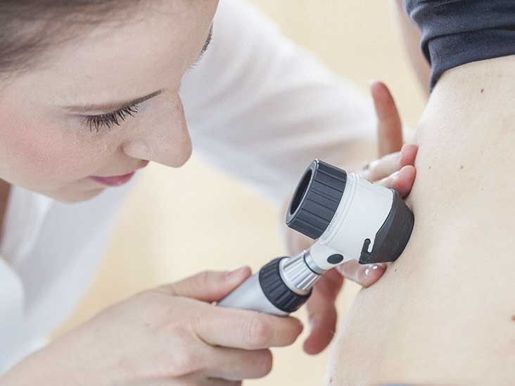
A dermatologist may first examine a suspicious spot with the naked eye and a dermatoscope, a handheld device that magnifies the skin and helps reveal pigment patterns that are difficult to see otherwise.
However, a visual exam alone cannot confirm melanoma. The only way to diagnose melanoma is through a biopsy, in which all or part of the suspicious lesion is removed and examined under a microscope by a pathologist.
What Your Pathology Report May Include
If melanoma is found, the pathology report may include details that guide treatment decisions:
- Breslow thickness: How deeply the melanoma has grown into the skin, measured in millimeters.
- Ulceration: Whether the skin over the melanoma has broken down.
- Margins: Whether melanoma cells are present at the edge of the biopsy sample.
- Mitotic activity: Information about how quickly melanoma cells appear to be dividing.
- Stage: A description of how advanced the melanoma is and whether it has spread.
Thickness matters because melanoma that has grown deeper generally carries a higher risk of spreading. This is one reason early detection is so valuable. A small-looking spot can still require attention, but catching melanoma before it grows deeply can dramatically simplify treatment.
Sentinel Lymph Node Biopsy
For some melanomas, especially those that are thicker or have higher-risk features, doctors may recommend a sentinel lymph node biopsy. The sentinel lymph node is the first lymph node most likely to receive drainage from the area where the melanoma started.
Testing that node can help determine whether melanoma cells have begun to spread beyond the original skin site. Not every person with superficial spreading melanoma needs this procedure. The decision depends on the pathology report, tumor thickness, ulceration, overall health, and other clinical factors.
Stages of Superficial Spreading Melanoma
Superficial spreading melanoma is staged in the same general way as other skin melanomas. The subtype describes its growth pattern, while the stage describes how far the cancer has progressed.
Stage 0: Melanoma In Situ
Stage 0 means melanoma cells are limited to the top layer of skin and have not invaded deeper tissue. This is often the earliest stage and is usually treated with surgical removal.
Stages I and II: Localized Melanoma
At these stages, melanoma has grown deeper into the skin but has not spread to nearby lymph nodes or distant organs. Surgery is often the main treatment, and some people may need additional testing depending on the tumor’s features.
Stage III: Regional Spread
Stage III melanoma has spread to nearby lymph nodes or nearby skin and lymphatic channels. Treatment may involve surgery, immunotherapy, targeted therapy, radiation in selected situations, or participation in a clinical trial.
Stage IV: Distant Spread
Stage IV melanoma has spread to distant organs or distant areas of the body. Treatment is more complex and may include immunotherapy, targeted drugs, surgery, radiation therapy, or clinical trials. Advanced melanoma treatment has improved significantly in recent years, especially through immune-based therapies and treatments directed at specific tumor mutations.
Superficial Spreading Melanoma Treatment Options
Treatment depends on the stage, depth, location, genetic features of the tumor, lymph-node involvement, and the person’s overall health. A dermatologist, surgical oncologist, medical oncologist, and other specialists may work together when melanoma requires more than a simple excision.
Surgery
Surgery is the main treatment for most early-stage superficial spreading melanomas. Doctors typically remove the melanoma along with a margin of normal-looking skin around it. This is called a wide local excision.
The amount of skin removed depends on the melanoma’s thickness and location. A small melanoma on the arm may leave a straightforward scar, while a lesion on the face, ear, hand, foot, or lower leg may require more specialized reconstruction or careful planning.
Immunotherapy
Immunotherapy helps the immune system recognize and attack cancer cells. These drugs are commonly used for higher-risk melanoma, melanoma that has spread to lymph nodes, or advanced melanoma.
Checkpoint inhibitor medicines are among the best-known immunotherapy options for melanoma. They can be highly effective for some people, although they can also trigger immune-related side effects affecting organs such as the skin, intestines, lungs, liver, thyroid, or joints. This is why treatment requires close monitoring.
Targeted Therapy
Some melanomas contain specific genetic mutations, including changes in the BRAF gene. When testing shows an actionable mutation, doctors may use targeted therapy drugs designed to interfere with cancer-growth signals.
Targeted therapy is not appropriate for every melanoma. It depends on the tumor’s molecular profile, stage, and treatment goals. This is a reminder that melanoma treatment is increasingly personalized; one person’s treatment plan may look very different from another person’s plan, even when both have the same melanoma subtype.
Radiation Therapy and Other Treatments
Radiation therapy may be used in selected cases, such as when melanoma involves certain lymph-node areas, returns locally, or spreads to specific organs. Surgery may also be used to remove isolated areas of disease in some patients.
Other treatment options may include intralesional therapy, systemic treatment combinations, or clinical trials. Traditional chemotherapy is used less often than in the past because modern immunotherapy and targeted therapy can offer better results for many advanced melanomas.
Can Superficial Spreading Melanoma Be Cured?
When superficial spreading melanoma is found early and removed before it spreads, treatment can be highly successful. Early-stage melanoma is often treated with surgery alone.
Once melanoma spreads to lymph nodes or distant organs, treatment becomes more involved. Even then, newer therapies have changed the outlook for many patients. The most important step is not trying to predict the future from an internet search at 1:17 a.m. It is getting accurate staging information and discussing your individual prognosis with a qualified oncology team.
Prevention: Reducing Your Melanoma Risk
No prevention strategy can eliminate melanoma risk completely, but protecting skin from UV damage can reduce risk and help prevent other skin cancers as well.
Smart Sun-Safety Habits
- Use broad-spectrum, water-resistant sunscreen with SPF 30 or higher.
- Apply sunscreen generously and reapply at least every two hours when outdoors.
- Wear wide-brimmed hats, sunglasses, long sleeves, and sun-protective clothing.
- Seek shade when UV exposure is strongest, especially around midday.
- Avoid tanning beds and sunlamps.
- Check the daily UV Index before spending extended time outdoors.
- Perform monthly skin self-exams.
- Schedule professional skin exams if you have many moles, atypical moles, prior skin cancer, or a strong family history of melanoma.
Sunscreen is important, but it is not a permission slip to spend all day roasting like a human rotisserie chicken. It works best as one part of a larger sun-protection plan that includes shade, clothing, and common sense.
When to See a Dermatologist
Make an appointment with a dermatologist promptly if you notice:
- A new mole or skin spot that looks unusual
- A mole that changes in shape, size, color, or texture
- A spot that bleeds, crusts, itches, or hurts repeatedly
- A lesion that does not heal
- An “ugly duckling” spot that looks different from your other moles
Do not try to remove a suspicious mole at home, burn it off with an over-the-counter product, or convince yourself that a photo from three years ago counts as medical surveillance. A timely exam and biopsy, when needed, are the safest way to get answers.
Experiences With Superficial Spreading Melanoma: What the Process Can Feel Like
The following section describes common real-world experiences people may encounter during evaluation and treatment. It is not a substitute for individualized medical advice, and every diagnosis and recovery process is different.
For many people, the experience starts with something surprisingly ordinary: a spouse notices a dark spot on the upper back, a person sees a changing mole after a shower, or someone compares vacation photos and realizes a freckle has become larger and more uneven.
The first emotion is often uncertainty rather than panic. People may wonder whether they are overreacting, whether the spot has “always looked like that,” or whether it can wait until a routine appointment months away. That hesitation is common, but a changing mole is worth checking. A dermatologist would rather reassure someone about a harmless mole than diagnose a melanoma later than necessary.
The dermatology visit itself is often faster than people expect. The clinician may examine the spot with a dermatoscope, compare it with the person’s other moles, and recommend a biopsy if the lesion has suspicious features. The word “biopsy” can sound intimidating, but it is usually a brief office procedure performed with local anesthesia. Many people say the anticipation was worse than the procedure itself.
Waiting for pathology results can be the hardest part. A person may replay every beach vacation, tanning-bed decision, sunburn, family history detail, and sunscreen mistake from the previous 20 years. This emotional spiral is understandable, but it is not especially helpful. The useful next step is to prepare questions: What type of melanoma is it? What is the Breslow thickness? Is there ulceration? Are the margins clear? Will I need more surgery or a sentinel lymph node biopsy?
When superficial spreading melanoma is diagnosed early, the treatment plan may be relatively straightforward: a wider excision, wound care, follow-up skin checks, and an adjustment period of learning to live with a scar. Even when the medical plan is simple, the emotional impact may not be. A small scar can become a large reminder, and some people become more anxious about every freckle afterward.
That anxiety can be managed by turning fear into a routine. Monthly self-exams, scheduled dermatology visits, photographs of moles, better sun habits, and conversations with loved ones can help restore a sense of control. Many people also find it useful to ask a partner or family member to check hard-to-see areas such as the scalp, back, buttocks, and backs of the legs.
For people with deeper tumors, lymph-node involvement, or advanced melanoma, the experience can involve more appointments, scans, oncology visits, treatment decisions, and side-effect management. Immunotherapy and targeted treatment may bring hope, but they can also bring fatigue, skin reactions, digestive symptoms, hormone changes, and the emotional stress of uncertainty.
Support matters. Some people lean on family, friends, cancer-support groups, therapists, faith communities, or melanoma survivor networks. Others prefer a small circle and a very organized spreadsheet. There is no correct emotional style for cancer. The goal is to stay informed, ask questions, report new symptoms, follow the treatment plan, and remember that a diagnosis is not a personality trait.
Superficial spreading melanoma can be serious, but it is also a condition where awareness can make a meaningful difference. A few minutes spent checking your skin may feel boring today. It can be incredibly important tomorrow.
Conclusion
Superficial spreading melanoma is the most common melanoma subtype, and it often begins as a changing or unusual-looking mole. Although its early outward growth pattern may make it appear less threatening, it can become dangerous if it grows deeper into the skin or spreads.
The best defense is early action: know the ABCDE warning signs, pay attention to new or evolving spots, avoid tanning beds, protect your skin from UV exposure, and schedule a dermatology appointment when something looks different. A suspicious mole is not always melanoma, but it always deserves a proper answer.