X-rays may be invisible, but they have a talent for making hidden things suddenly very obvious. A cracked wrist, a swallowed coin, a suspicious shadow on a lung, a cavity hiding between teeth, even the hot drama around exploding stars in spaceX-rays have a way of showing up where ordinary eyesight politely gives up.
Since their discovery in 1895, X-rays have changed medicine, dentistry, astronomy, airport security, art conservation, engineering, and science education. They are one of those rare inventions that feel both old-fashioned and futuristic at the same time. We associate them with hospital rooms and lead-lined doors, yet they also help scientists study black holes, inspect airplane parts, and examine priceless paintings without peeling off a single brushstroke.
Below are nine transparently amazing facts about X-rays, written for curious readers who like their science accurate, useful, and just a little less stiff than a hospital waiting-room chair.
1. X-Rays Were Discovered by Accident
The story of X-rays begins with a beautiful scientific accident. In 1895, German physicist Wilhelm Conrad Röntgen was experimenting with cathode rays in a dark laboratory. He noticed that a nearby fluorescent screen began to glow even though the equipment was covered. Something invisible was passing through the barrier.
Röntgen did not know what the mysterious rays were, so he called them “X-rays,” using the letter X the way mathematicians use it: for something unknown. The name stuck. It sounds cooler than “invisible body-peeking energy beams,” which is probably for the best.
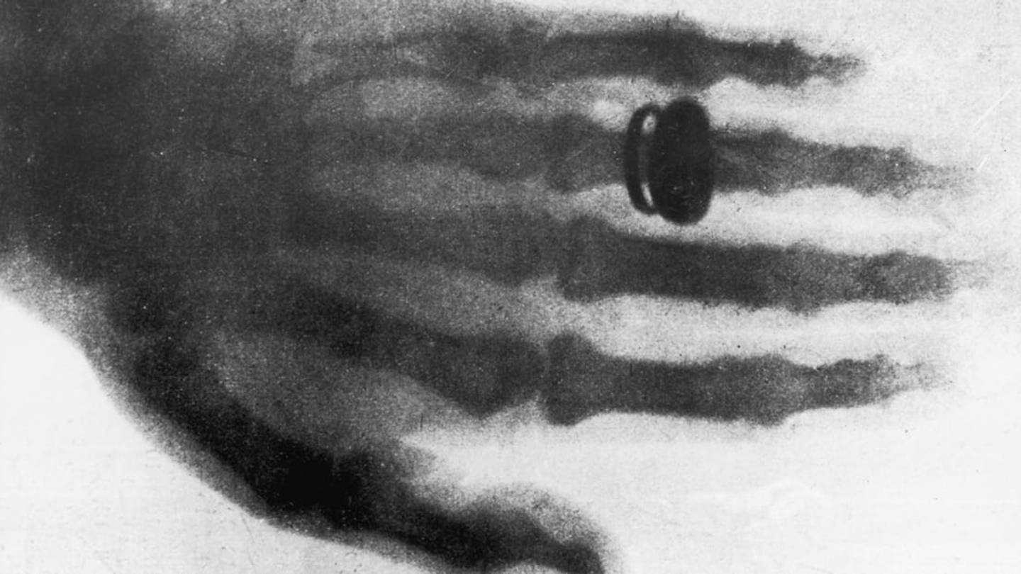
One of the earliest and most famous X-ray images was of his wife Anna Bertha Ludwig’s hand. The image showed the bones of her fingers and her wedding ring. Imagine seeing your own skeleton while still being very much alive. According to historical accounts, she was understandably startled. Science had just handed humanity a new superpower: the ability to see inside the body without opening it.
2. X-Rays Are a Form of LightJust Not the Kind Your Eyes Can See
X-rays are part of the electromagnetic spectrum, the same family that includes radio waves, microwaves, infrared light, visible light, ultraviolet light, and gamma rays. The difference is energy and wavelength. X-rays have much higher energy and much shorter wavelengths than visible light.
Your eyes are built to detect only a tiny slice of the electromagnetic spectrum. X-rays sit far outside that visible range, so we cannot see them directly. Instead, X-ray machines use detectors, film, or digital sensors to capture how these rays pass through materials.
That is why an X-ray image is not really a photograph in the regular sense. It is more like a shadow map. The image shows how much radiation passed through one part of the body compared with another. Dense materials block more X-rays; softer materials let more pass through.
3. X-Rays Do Not “See Through Everything”
Popular culture often treats X-ray vision like a magical ability to see through anything. Real X-rays are more selective. They pass easily through some materials but are absorbed or blocked by others.
Air absorbs very little X-ray radiation, so lungs often appear dark on a chest X-ray. Soft tissues, such as muscles and organs, appear in shades of gray. Bones absorb more X-rays because they contain calcium, so they usually appear white or light. Metal objects, such as dental fillings, surgical hardware, jewelry, and bullets, can appear very bright because they block a lot of radiation.
This difference in absorption is what makes X-rays so useful. A broken bone stands out because the dense bone produces a strong contrast against surrounding tissue. A dentist can spot decay because damaged areas of a tooth may absorb radiation differently. A doctor can examine the lungs because air-filled spaces and surrounding tissues create visible patterns.
4. There Is More Than One Kind of Medical X-Ray
When people say “I got an X-ray,” they often mean a standard radiograph, such as a chest X-ray or hand X-ray. But X-ray technology comes in several forms.
Radiography
Radiography creates still images. It is commonly used for bones, teeth, chest exams, and certain abdominal images. It is quick, widely available, and often one of the first imaging tools used after an injury.
Computed Tomography
Computed tomography, better known as CT, uses X-rays and computer processing to create cross-sectional images of the body. Instead of one flat shadow image, CT can produce detailed slices and 3D-style views. CT scans can be especially helpful for trauma, internal bleeding, tumors, blood clots, and complex anatomy.
Fluoroscopy
Fluoroscopy is sometimes described as a moving X-ray. It lets healthcare providers watch body structures in real time. It can help guide procedures, examine the digestive tract, place medical devices, or assist with joint and spine treatments.
Mammography
Mammography is a specialized X-ray exam designed to image breast tissue. It plays an important role in breast cancer screening and diagnosis.
So yes, “X-ray” is not just one thing. It is a whole toolbox, and doctors choose the tool based on the medical question they need to answer.
5. X-Rays Help With Much More Than Broken Bones
Broken bones are the celebrity use case for X-rays, but they are not the whole show. X-rays are used to detect pneumonia, examine heart size, locate swallowed objects, evaluate arthritis, guide dental treatment, monitor certain medical devices, and help diagnose conditions involving the chest, abdomen, spine, joints, and teeth.
Outside medicine, X-rays also have impressive side gigs. Airport security systems use X-ray imaging to inspect luggage. Engineers use industrial X-rays to check welds, pipelines, aircraft components, and manufactured parts for hidden cracks or flaws. Museums and art conservators use X-rays to look beneath the surface of paintings, sometimes revealing earlier sketches, repairs, or even completely different images hidden under the final artwork.
In other words, X-rays are not just hospital technology. They are professional secret-finders. If something important is hidden behind a surface, there is a decent chance X-rays have been invited to investigate.
6. X-Rays Use Ionizing Radiation, So Safety Matters
X-rays are powerful because they are ionizing radiation. That means they carry enough energy to remove electrons from atoms. This ability is exactly what makes them useful for imaging, but it also means unnecessary exposure should be avoided.
For most standard diagnostic X-rays, the radiation dose is low, and the medical benefit often outweighs the small risk when the exam is necessary. Still, healthcare teams follow a safety principle often called ALARA: “as low as reasonably achievable.” The goal is to use the smallest radiation dose needed to get a useful image.
Modern imaging systems are designed to be more efficient than older technology. Digital detectors, focused beams, better positioning, and improved software can help reduce repeat images and unnecessary exposure. But safety still depends on asking the right question: Is this exam needed, and will it help guide care?
A good X-ray is not the one taken “just because.” It is the one that answers a real clinical question.
7. Children and Pregnancy Require Extra Thought
X-ray imaging can be appropriate for children and pregnant patients when medically necessary, but healthcare providers are especially careful in these situations. Children are more sensitive to radiation than adults, and they have more years ahead of them during which long-term effects could theoretically develop. That is why pediatric imaging often uses child-sized settings rather than adult settings.
Pregnancy also requires careful decision-making. If an imaging exam does not involve the abdomen or pelvis, the exposure to the fetus may be very low. However, healthcare providers still consider whether ultrasound, MRI, delayed imaging, or modified technique would be better depending on the situation.
This does not mean patients should panic if an X-ray is recommended. It means they should communicate. If you are pregnant, might be pregnant, or are caring for a child who needs imaging, tell the healthcare team. The safest imaging decision is usually the one made with complete information.
8. Dental X-Ray Safety Has Changed With Technology
Dental X-rays are one of the most familiar uses of X-ray imaging. They help dentists find cavities between teeth, check bone levels, monitor tooth development, examine roots, and plan treatments that cannot be fully evaluated by looking inside the mouth with a mirror and heroic optimism.
Modern dental radiography has become more targeted and efficient. Digital sensors can reduce the need for higher exposure compared with older film methods, and better beam collimation helps focus the X-rays on the area being examined.
One interesting recent shift is the guidance around lead aprons and thyroid collars in dentistry. For many years, patients expected a heavy lead apron before dental X-rays. Updated professional recommendations now emphasize that shielding is not always needed for dental imaging and can sometimes interfere with the image, potentially causing retakes. The bigger safety win is making sure the X-ray is necessary, properly aimed, and done correctly the first time.
That may feel surprising if you grew up thinking the apron was the main event. But in modern imaging, precision often protects better than tradition.
9. X-Rays Let Us Study the Universe
X-rays are not only made by machines in clinics. They are also produced by extremely energetic events in space. Hot gas around black holes, neutron stars, supernova remnants, and galaxy clusters can emit X-rays. The universe, it turns out, is not quiet. It is basically running a cosmic light show, but most of it is invisible to human eyes.
Earth’s atmosphere blocks most incoming cosmic X-rays, which is excellent for life but inconvenient for astronomers standing on the ground with telescopes. To study X-ray sources in space, scientists use satellites and space telescopes that orbit above the atmosphere.
X-ray astronomy has helped scientists understand some of the most extreme objects in the universe. It reveals hot, violent, high-energy regions that visible-light telescopes cannot fully capture. So the same type of radiation that helps doctors find a fracture can also help researchers study black holes. That is a pretty strong résumé.
Real-Life Experiences With X-Rays: What People Usually Notice
For many people, the first memorable X-ray experience happens after a fall, a sports injury, a dental checkup, or a mysterious cough that refuses to leave politely. The process is usually faster and less dramatic than people expect. You may walk into an imaging room imagining a science-fiction chamber, only to find a table, a machine, a friendly technologist, and instructions like “hold still” or “take a deep breath.” The hardest part is often not the X-ray itself but staying in an awkward position while pretending you are totally comfortable.
A typical bone X-ray may take only a few minutes. The technologist positions the body part, steps behind a barrier or out of the room, and captures the image. You do not feel the X-rays. There is no zap, buzz, heat, or superhero origin story. If the injured area hurts, positioning may be uncomfortable, but the imaging itself is painless.
Dental X-rays are a different kind of tiny adventure. Many patients remember biting down on a small sensor while trying not to drool, move, or question every life choice that led to that moment. Bitewing images can feel awkward, especially for people with a sensitive gag reflex, but they are usually over quickly. The reward is practical: dentists can see problems hiding between teeth long before those problems become expensive little disasters.
Chest X-rays feel even more routine. You may stand against a flat detector, roll your shoulders forward, hold your breath for a second, and then you are done. In emergency settings, portable X-ray machines can come directly to the bedside, which is especially helpful for patients who cannot easily move.
CT scans feel more high-tech because the scanner looks like a large ring. The table moves through the opening while the machine captures multiple X-ray images. Some CT exams use contrast material, which may create a warm sensation or a metallic taste. That part surprises people, but medical staff usually explain it beforehand.
The biggest emotional experience around X-rays is often waiting for results. The image may be captured quickly, but patients naturally want answers right away. Is it broken? Is the lung clear? Is that tooth doomed? In urgent situations, results may come quickly. In routine cases, a radiologist or qualified clinician reviews the images and sends a report to the ordering provider.
The best patient experience comes from communication. Ask why the X-ray is needed, what it can show, whether alternatives exist, and when results will be available. These questions are not annoying; they are part of being an informed patient. X-rays may be famous for seeing through things, but patients should never feel left in the dark.
Conclusion: X-Rays Make the Invisible Useful
X-rays are one of the most important discoveries in modern science because they turned invisibility into information. They help doctors diagnose injuries, dentists catch hidden decay, engineers inspect critical structures, conservators study artwork, and astronomers explore violent cosmic events far beyond Earth.
They are not magic, and they are not risk-free. They work because of real physics, careful technique, and thoughtful safety standards. Used appropriately, X-rays offer a remarkable balance: a brief exposure that can provide life-changing information.
More than a century after Röntgen’s unexpected laboratory glow, X-rays remain transparently amazing. They remind us that some of the most powerful discoveries begin with someone noticing that something strange is happeningand being curious enough to find out why.